Figure S4.
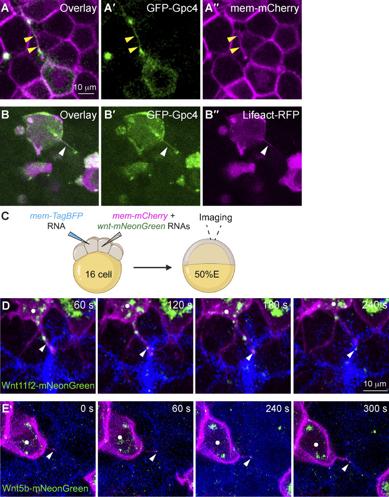
Actin-based filopodia deliver Wnt proteins to neighboring cells. (A–A′′) Snapshots from confocal time-lapse imaging of zebrafish blastula cells (labeled with mem-mCherry, in magenta) showing a GFP-Gpc4 expressing cell extending a long cellular protrusion (yellow arrowheads). (B-B′′) Snapshots from confocal time-lapse imaging of zebrafish blastula cells, showing a GFP-Gpc4-labeled protrusion (white arrowheads) that is colabeled Lifeact-RFP (magenta). (C) Schematic diagram illustrating mosaic injection, with distinct cells of embryos at the 16-cell stage injected with specific sets of RNAs, as indicated. Confocal live imaging was performed, with a focus on the regions where the two populations of labeled cells were in close proximity. (D and E) Snapshots from confocal time-lapse imaging (Video 5) showing that mem-mCherry labeling protrusions extended from Wnts-mNeonGreen–expressing cells (white dots) transport Wnt11f2-mNeonGreen (D) or Wnt5b-mNeonGreen (E) to the neighboring BFP-expressing cells (Video 5). White arrowheads, Wnt-expressing puncta on protrusions.

