Figure S6.
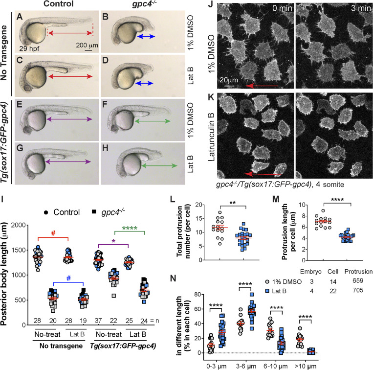
Inhibition of actin polymerization by Lat B blocks the rescue mediated by endodermal expression of GFP-Gpc4. (A–H) Bright-field images of the indicated embryos. Lines with double arrows indicate length of the posterior body axis; lines of the same color are equal in length. (I) Average posterior body length in embryos shown in A–H, from three independent experiments (represented by different color symbols), with the number of embryos indicated. Colors of the P values correspond to the embryos in which the posterior body is marked with lines of the same color. (J and K) Snapshots from confocal time-lapse imaging performed on gpc4−/−/Tg(sox17:GFP-gpc4) embryos treated with 1% DMSO and Lat B (0.15 µg/ml; Video 9). Red arrows, direction of migration of the endodermal cells. (L–N) The total number of protrusions (L), the length of the protrusion (M), and the percentages of protrusions of different lengths (grouped into 3-µm bins; N) in each endodermal cell. The number of embryos, cells, and protrusions analyzed is indicated. Data are mean ± SEM. #, P > 0.05; *, P < 0.05; **, P < 0.01, ****, P < 0.0001; unpaired Student's t test.

