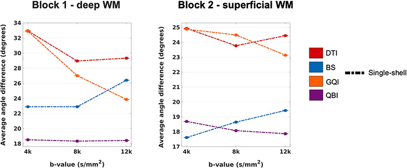
Fig. 15. Effect of b-value on the accuracy of dMRI orientation estimates.
The plots show the absolute angular error, averaged over all included WM voxels from all PSOCT sections in each sample, as a function of b-value for a single-shell sampling scheme, at 1 mm spatial resolution. Line colors represent different orientation reconstruction methods.

