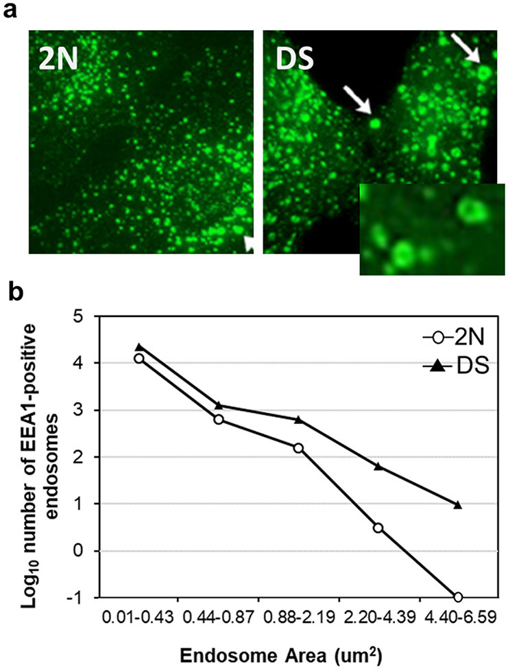
Fig.3. Early endosomes in DS.
a, Representative immunofluorescence micrographs of rab5-positive endosomes in DS and age-matched 2N controls. DS fibroblast show a striking enlargement in the size of rab5-positive endosomes compared to 2N. b, The numbers of EEA1-positive early endosomes in all size groups are also increased in the DS fibroblasts compared to 2N controls (total fibroblasts examined: 2N fibroblasts = 80; DS fibroblasts = 80).

