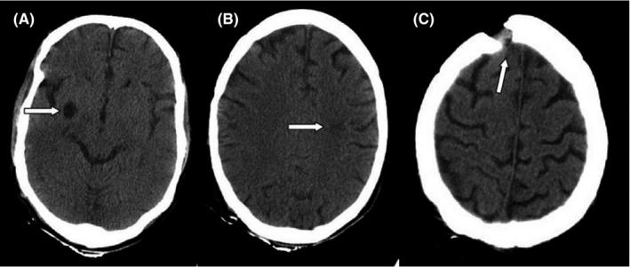
FIGURE 1.

Computed tomography (CT) scans of the brain of the patient. A hypodensity in the left peritoneal (A), CSF‐like density in the right basal ganglia (B), a bone defect in the right frontal (C)

Computed tomography (CT) scans of the brain of the patient. A hypodensity in the left peritoneal (A), CSF‐like density in the right basal ganglia (B), a bone defect in the right frontal (C)