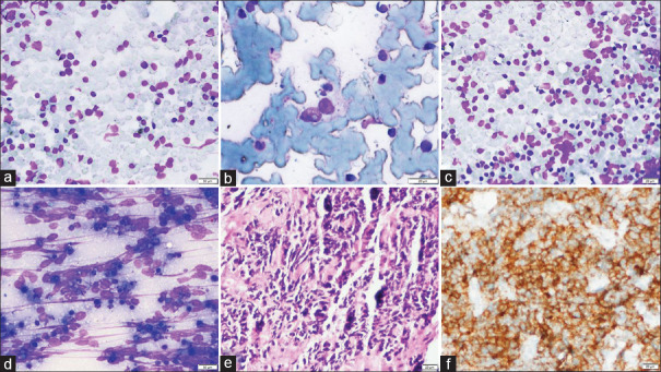
Figure 2.
Category 1 (a–c): FNA cytology smears (case no. 14) showing predominantly large germinal center cells mimicking atypical lymphoid cells (a; MGG, 40X), binucleated RS-like cell (case no. 4) (b; MGG, 200X), and sheets of large monomorphic cells mimicking low-grade lymphoma (case no. 11) (c; MGG, 40X); Category 2 (d–f): FNA cytology smears (case no. 1) shows singly scattered atypical cells with marked crushing artifact (a; MGG, 200X), histopathology shows sheets of tumor cells (b; H and E, 100X) which are positive for CD56 immunostain confirming a small cell carcinoma (c; CD56, 100X)

