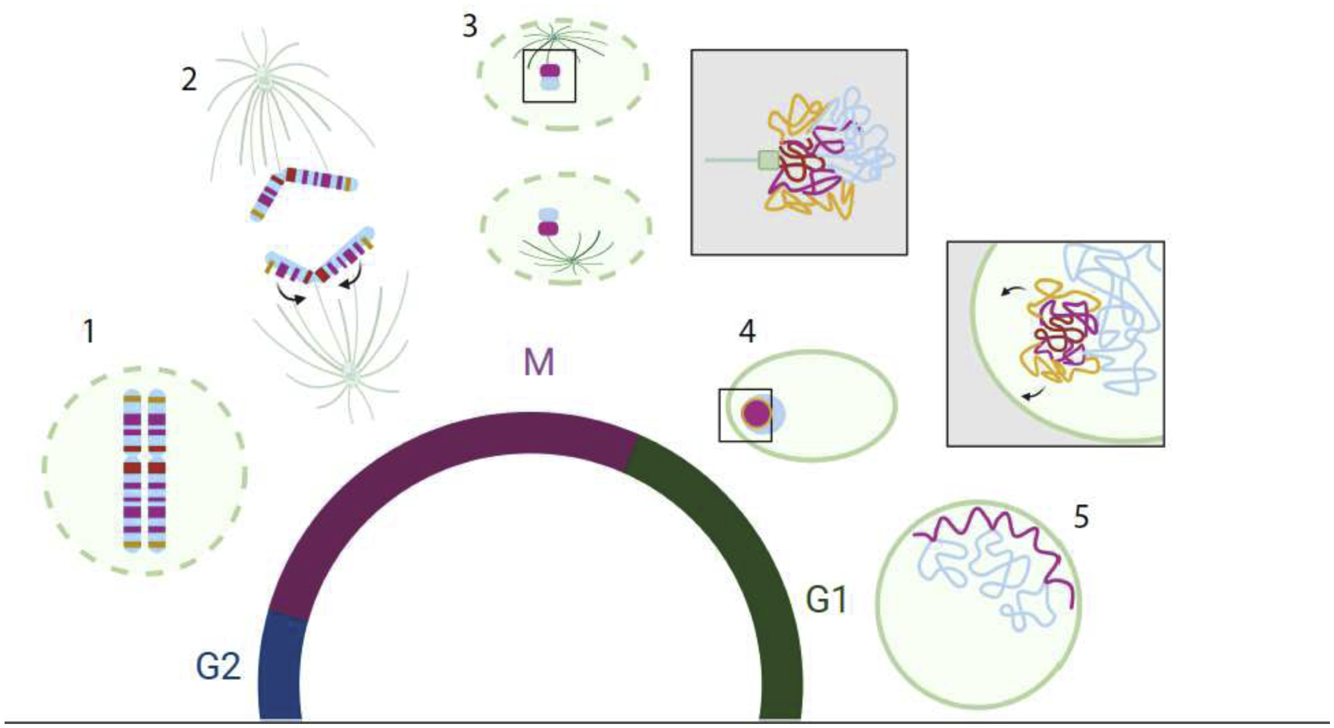
Figure 3: Broad view of genome organization through mitosis.

A schematic representing the progression of genome organization through mitosis and re-establishment. Lamins and spindles (green) centromere (dark blue) Centromere proximal LADS (red) telomere proximal LADs (Orange) LADs (pink) nonLADs (light blue). Clockwise from bottom left: 1. (M) As cells enter mitosis the nuclear envelope breaks down (green dashed line) and nuclear lamins become phosphorylated and disperse into the cytoplasm. At this time interphase genome organization is also dismantled and chromosomes become configured into a linearly arranged looping structure. 2. As cells proceed into anaphase and sister chromosomes begin to segregate the LAD regions begin to coalesce into an intrachromosomal cluster towards the centromere (arrows), excluding nonLAD regions, thus separating LADs/nonLADs (A/B compartment). Telomeric proximal LADs are shown in yellow. 3. As the nuclear envelope and lamina reform the LAD regions remain in tight agglomerates and separated from nonLAD regions. 4. (G1) As cells enter early G1 the nonLAD regions (A-comaprtment) begin to form looping structures and the LAD agglomerates increasingly associate with the nuclear lamina where they eventually become constrained. Possibly due to the orientation of the chromosomes when LAD coalesce, the telomere proximal LADs make contact with the nuclear envelope first followed later by centromere proximal LADs. 5. As G1 continues, A/B compartmentalization strengthens, with increased inter-chromosomal contact within the A-compartment, and increased interaction (flattening) of LADs with the nuclear lamina. Black boxes indicate magnified views.
