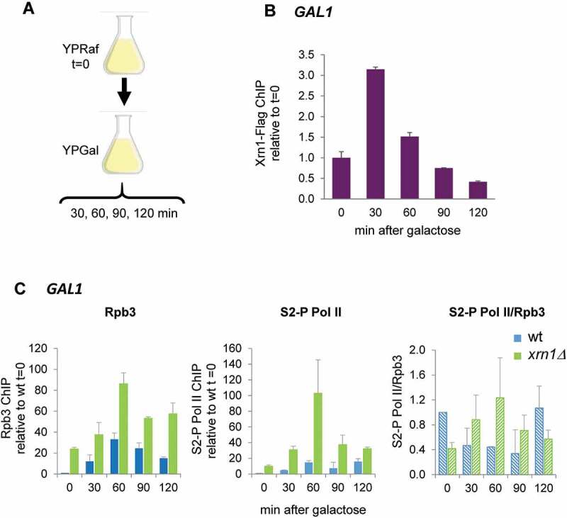
Figure 7.

Xrn1 binds to GAL1 upon activation and prevents the accumulation of RNAPII. (A) Scheme of the assay. Exponentially growing cells in raffinose (YPRaf) medium were washed and transferred to galactose medium (YPGal), and samples were collected at the indicated times. (B) Chromatin immunoprecipitation (ChIP) analysis of Xrn1-Flag was made using anti-Flag antibodies and binding to GAL1 ORF was determined. (C) ChIP analysis of RNAPII binding at GAL1 ORF in wild type (wt) and xrn1Δ cells. RNAPII was pulled down with anti-Rpb3p (left panel) and anti-Rpb1-CTDSer2-P (middle panel) antibodies in two aliquots of the same whole cell extract sample. Ratio between Rpb1-CTDSer2-P (S2-P Pol II) and Rpb3 ChIP is shown (right panel). ChIP data were calculated and normalized as described in Figure 5. Mean and standard deviation from three independent experiments are shown
