Figure 3.

Validation the DTYMK expression in HCC cell lines and HCC patients
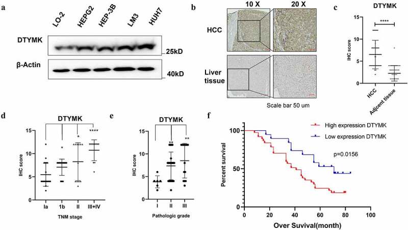
(a) Western blot analysis of DTYMK in HCC cells including LO-2, HEPG2, HEP3B, LM-3, and Huh-7. β-Actin was used as a loading control; (b) expression of DTYMK protein by IHC. DTYMK protein was predominantly detected at the cytoplasm. Arepresentative case shows the upregulation of DTYMK protein in HCC compared to that in its adjacent normal liver tissue; (c) semi-quantitative analyses of IHC staining of DTYMK in 60 HCC patients TMAs with tumor sample and adjacent tissue; (D) semi-quantitative analyses of IHC staining of DTYMK in 60 HCC patients TMAs with different TNM-stage patient; (e) semi-quantitative analyses of IHC staining of DTYMK in 60 HCC patients TMAs with different pathology grade patients (f) Kaplan–Meier of survival of 60 patients with HCC (two groups stratified by DTYMK expression level. Differences between the groups were shown by a log-rank test.)
