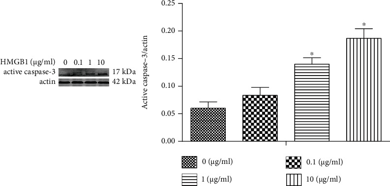
Figure 7.

HK-2 apoptosis is increased once exposed to HMGB1. The HK-2 cells were challenged by HMGB1 at various concentrations (0, 0.1, 1, and 10 μg/mL) for 24 h. Representative Western blotting analysis of active caspase-3 expression in different groups. ∗P < 0.05 compared with the control group.
