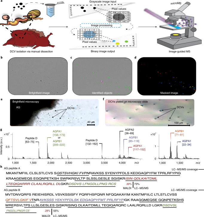
Fig. 1. High-throughput workflow for label-free single-DCV targeting and MS analysis.
a, Schematic of MALDI MS workflow for high-throughput single-DCV measurements. b, Brightfield image of DCVs distributed on a glass slide. c, Identification of primary objects (DCVs) using image-processing software. d, Masked image output of identified DCVs. The colored spots represent primary objects (DCVs) recognized as ‘foreground’ and are marked with a maximal pixel intensity value. Anything not identified as an object is treated as ‘background’ and is set to a zero-pixel intensity value. e, Brightfield image of mechanically induced DCV release from the AG. f, DCVs plated on a glass slide for relative DCV density estimation using brightfield microscopy. Each slide held DCVs from three animals (biological replicates) and a total of three slides (technical replicates) were prepared, where 598 DCVs were measured. g, Mass spectra demonstrating the coverage of AG peptides detected in single-DCV measurements. h, AGPA1 (XP_012945143.1) and AGPB1 (XP_012945142.1) prohormone sequences with corresponding MALDI MS-detected peptides italicized and font colored to match the annotated spectra in g. AG peptide assignments were validated using LC–MS/MS and performed on AG extracts (n = 3). The peptides detected by LC–MS/MS are underlined in black. a.u., arbitrary units.

