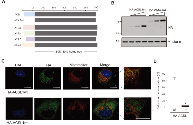
Fig. 1. Structure of ACSLs and the ACSL1 mutant.
(A) Sequence comparison of mouse ACSL family members and the N-terminal-deleted ACSL1 mutant (ACSL1mt). (B) ACSL1 expression was detected by western blot analysis after transfection of pc-HA-ACSL1 wild type (wt) and pc-HA-ACSL1 mutant (mt) into COS7 cells. (C) Immunofluorescence micrographs after transfection of pc-HA-ACSL1wt or pc-HA-ACSL1mt in COS7 cells. Scale bars = 10 μm. (D) The ratio of mitochondrial co-localized HA-ACSL1 to total HA-ACSL1 was measured. n = 4, *P < 0.05 vs HA-ACSL1wt, by t-test.

