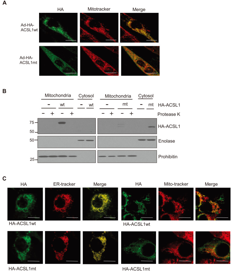
Fig. 2. Intracellular distribution of wild type ACSL1 (ACSL1wt) and mutant ACSL1 (ACSL1mt) in C2C12 myotubes.
(A) Immunofluorescence micrographic analysis was performed after C2C12 myotubes were infected with Ad-HA-ACSL1wt or Ad-HA-ACSL1mt. Scale bars = 10 μm. (B) Subcellular fractionation was performed after infection with Ad-HA-ACSL1wt or Ad-HA-ACSL1mt into C2C12 myotubes. One half of the mitochondrial fraction was treated with protease K. Mitochondrial fractionation was confirmed by western blotting with antibodies against enolase (cytosol) and prohibitin (mitochondria). (C) Immunofluorescence micrographic analysis was performed after HepG2 cells were infected with Ad-HA-ACSL1wt or Ad-HA-ACSL1mt. Scale bars = 10 μm.

