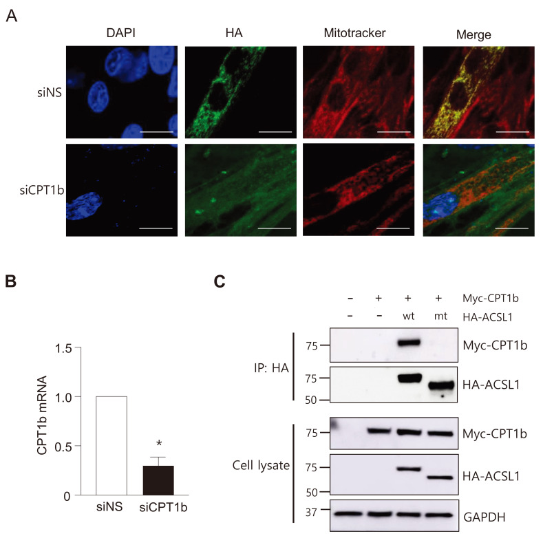
Fig. 3. ACSL1 interacts with a mitochondrial protein CPT1b in myotubes.
(A) Immunofluorescence micrographs were conducted after C2C12 myotubes were treated with siNS (nonspecific) or siCPT1b for 48h. Scale bars = 10 μm. (B) Relative mRNA levels of CPT1b after treatment with siCPT1b in myotubes. n = 3, *P < 0.05 vs control, by t-test. (C) COS7 cells were co-transfected with pc-HA-ACSL1wt or pc-HA-ACSL1mt and pCMV-CPT1b-Myc. Immunoprecipitation (IP) was conducted with an HA antibody, and then western blotting was performed with HA or Myc antibodies.

