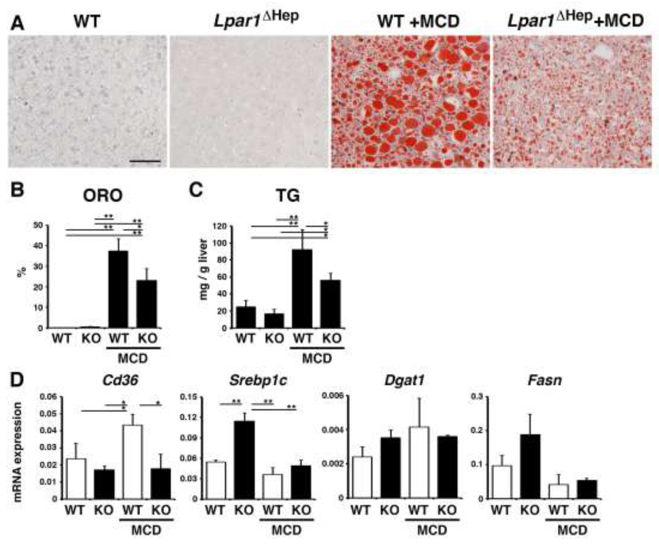
Fig. 5. Hepatocyte-specific deletion of Lpar1 gene reduces steatosis in mice fed the MCD diet.

Control Lpar1flox/flox (WT) and Lpar1ΔHep (KO) mice were fed with the MCD diet for 10 days (male, n=3 for each group). (A) Oil red O staining of the liver tissues from the WT and Lpar1ΔHep mice fed with the control or MCD diet. After the MCD diet, the Lpar1ΔHep liver shows less accumulation of lipids compared to the WT liver. (B) Quantification of oil red O (ORO) staining in each group. (C) Measurement of triglyceride levels (TG) in the livers. (D) mRNA expression of the liver tissues was analyzed by QPCR. mRNA expression values were normalized against Gapdh. Each value is the mean ± SD of triplicate measurements. *, p < 0.05; **, p < 0.01.
