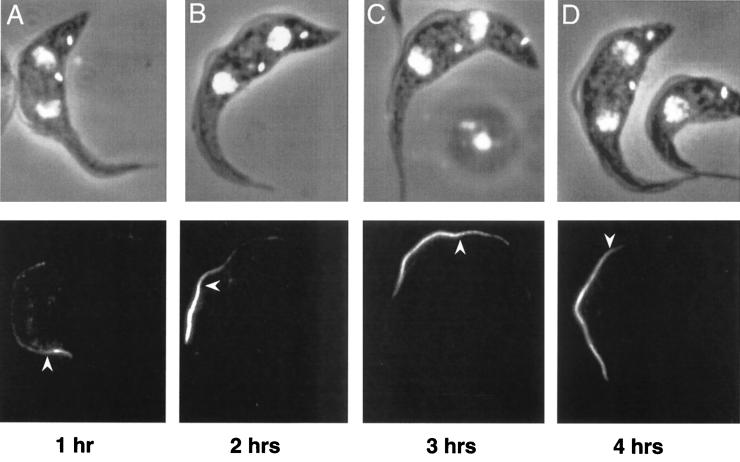
FIG. 4.
Pattern and timing of PFR assembly. PFRAtag trypanosomes were grown for 1 (A), 2 (B), 3 (C) or 4 h (D) in the presence of tetracycline and were analyzed by immunofluorescence with the antitag BB2 monoclonal antibody. The top panels show the DAPI images (white) merged to the phase contrast images, and the bottom panels show the immunofluorescence signal. The arrowhead indicates the break point between the bright distal and the less intense proximal staining.

