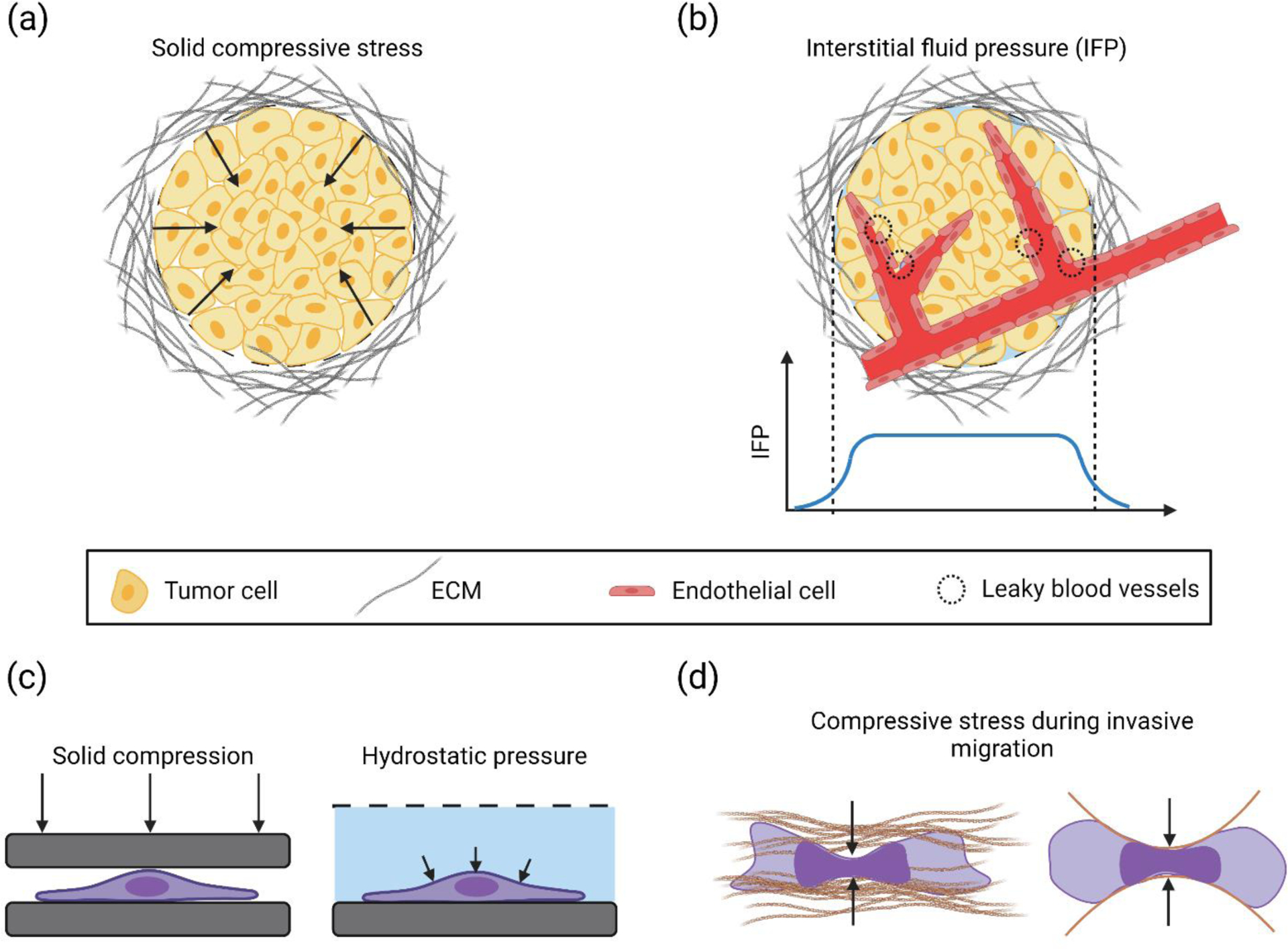
Figure 1. Build-up of solid compressive stress or hydrostatic pressure in tumors, and in vitro assays to study them.

(a) A solid tumor mass surrounded by dense ECM. Overcrowding of cells in the tumor microenvironment due to abnormal cell proliferation displaces the surrounding ECM and causes a buildup of solid compressive stress (black arrows (b) Leaky/permeable blood vessels in the solid tumor cause plasma leakage which, combined with a lack of functioning lymphatic vessels, leads to elevated interstitial fluid pressure (IFP) in the bulk of the tumor, with a gradient near the tumor periphery. The IFP distribution in the tumor mass is shown by the blue curve. (c) In vitro approaches to apply solid compressive stress and hydrostatic pressure (black arrows) on cancer cells. (d) Solid compressive stress acts on an invading cancer cell in the confined environment of the extracellular matrix. Right image shows a schematic of a cancer cell migrating through microfabricated confining channels. Compressive stresses are indicated by solid black arrows.
