Figure 3.
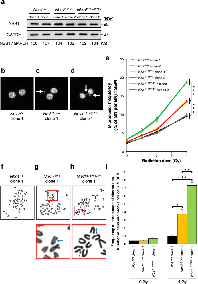
Radiation-induced chromosomal instability of Nbs1 I171V edited MEF clones. (a) Western blotting analysis data showing the expression levels of Nbs1 protein in Nbs1 I171V edited MEF clones. Full-length gel is presented in Supplementary Fig. 5. The GAPDH antibody was used as a loading control. The intensity of Nbs1 bands was normalized to that of GAPDH and shown as a percentage, regarding the score of Nbs1+/+ clone 1 as 100%. (b–d) Metafer MN Search images showing the cytokinesis-blocked Nbs1 I171V edited MEFs stained with DAPI. Arrowheads indicate MN. Nbs1+/+-BN cell without MN (b), Nbs1I171V/+-BN cell with one MN (c), Nbs1I171V/I171V-BN cell with three MN (d). (e) Percentage of IR-induced MN formation in Nbs1 I171V edited MEF clones (mean ± SEM; t-test; n = 3; > 1000 BN cells). (f, g) Representative metaphase of Nbs1 I171V-edited MEFs after 4 Gy irradiation. Remarkable aberrations are enlarged. Arrows indicate chromosomal breakages. (i) Frequency of IR-induced chromosomal aberrations in Nbs1 I171V edited MEF clones (mean ± SEM; t-test; n = 3; > 50 cells).

