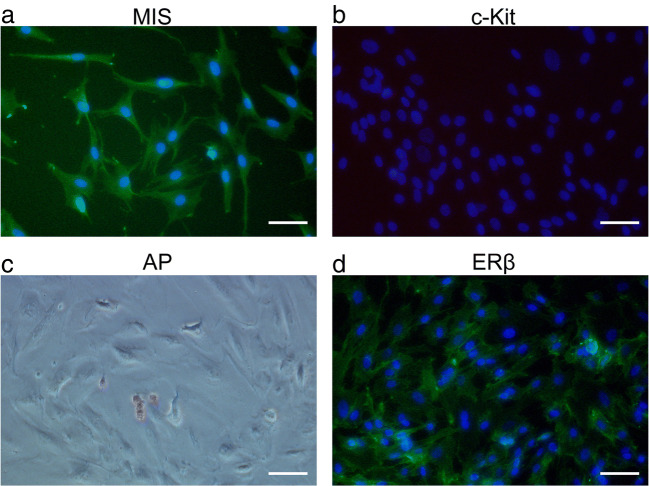
Fig. 1.
Identification of primary Sertoli cells isolated from human fetal testes. Primary human Sertoli cells were isolated from 16 human fetal testes. Immunocytochemical staining was performed on cells passaged no more than 12 times and prior to confluency. Representative images are shown. The nuclei are stained with DAPI (blue). Scale bar: 50 μm. a Immunostaining for Mullerian inhibiting substance (MIS) (green), a specific Sertoli cell marker; b immunostaining for c-Kit (green), a specific germ cell marker; c immunostaining for alkaline phosphatase (AP) (red), a stem cell marker; and d immunostaining for estrogen-responsive (ER)β (green)

