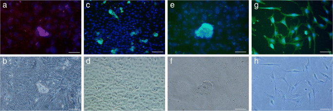
Fig. 4.
Identification of fetal SSCs and Sertoli cells isolated from human fetal testes. Immunocytochemical staining was performed in a unique co-culture system including human fetal SSCs and Sertoli cells. a Immunostaining of c-Kit (green); b phase-contrast microscopic image of a; c immunostaining of SSEA-4 (green); d phase-contrast microscopic image of c; e immunostaining of Oct-4 (green); f phase-contrast microscopic image of e; g immunostaining of MIS (green); and h phase-contrast microscopic image of g. The nuclei are stained with DAPI (blue). Scale bar: 50 μm

