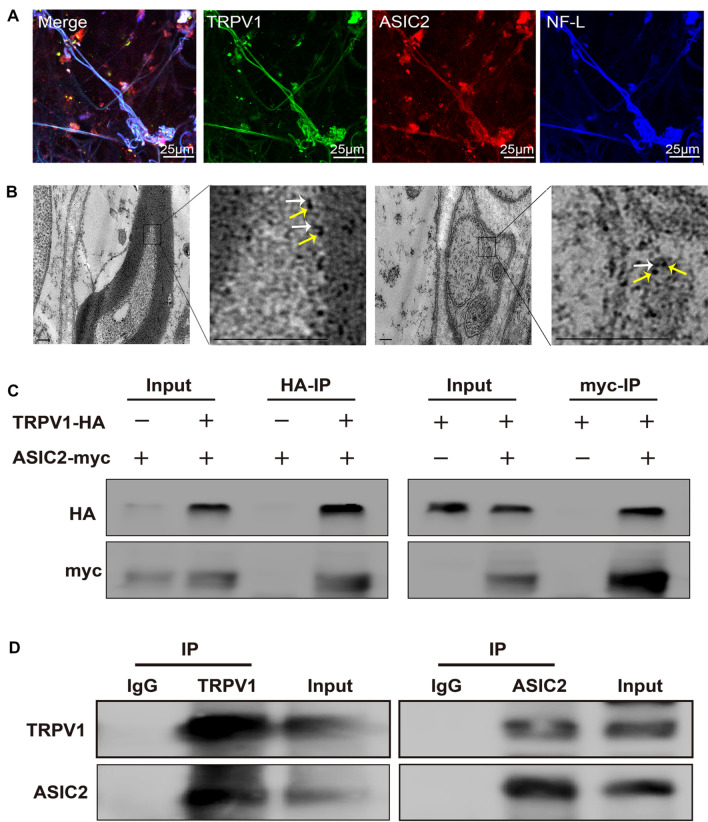
Fig. 5.
ASIC2 interacts with TRPV1 by forming a compact complex in baroreceptors. A Representative images showing the immunoreactivity of ASIC2 (red), TRPV1 (green), and the neurofilament marker NF-L (blue) in baroreceptor terminals within the aortic arch adventitia. B Representative immunoelectron microscopic images of myelinated and unmyelinated nerve fibers from rat aortic baroreceptor nerve terminals. White arrows mark the 4-nm colloidal gold particles labelling ASIC2 ion channels. Yellow arrows mark 18-nm colloidal gold particles labelling TRPV1 ion channels. Scale bar, 200 nm. C Co-immunoprecipitation (IP) experiments carried out on lysates of HEK293T cells expressing ASIC2-myc and/or TRPV1-HA. The transfected constructs are indicated above. Anti-HA (HA-IP, left) or anti-myc (myc-IP, right) antibodies were used for IP. The antibodies used for western blotting (WB) analysis are shown on the left. D Co-IP experiments carried out on rat aortic arch baroreceptor tissue lysates. Left: ASIC2 and TRPV1 detected by WB analysis of immunoprecipitate collected with anti-TRPV1 antibodies (TRPV1-IP). Right: TRPV1 and ASIC2 detected by WB in immunoprecipitate collected with anti-ASIC2 antibodies (ASIC2-IP).

