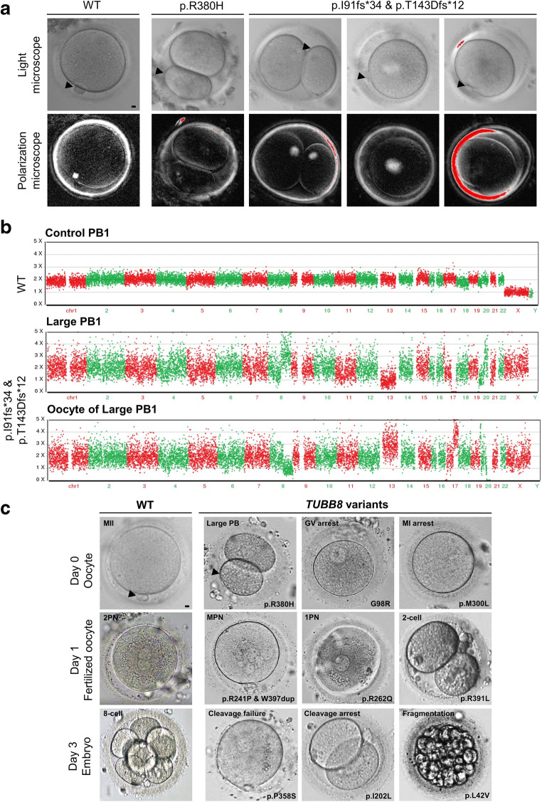
Fig. 2.
Morphology of oocytes, zygotes, and embryos from control individuals and patients. a PB1 oocytes from a control and patients in families 1 and 2 were examined by light and polarization microscopy. The control oocyte has a visible spindle located near PB1, while it is only very weakly visible in the R380H variant. No detectable spindle can be observed in the p.I91Hfs*34 and p.T143Dfs*12 compound heterozygous variants. Scale bar = 10 μm. b Control PB1 has a diploid chromosome copy number. The large PB and oocyte had abnormal chromosome compositions, including partial chromosome trisomy and partial chromosome monosomy. c Morphology of a control and an affected individuals’ oocytes on day 0, zygotes on day 1, and embryos on day 3 after fertilization presenting various morphological abnormalities. Arrow indicated the PB1. Scale bar = 10 μm

