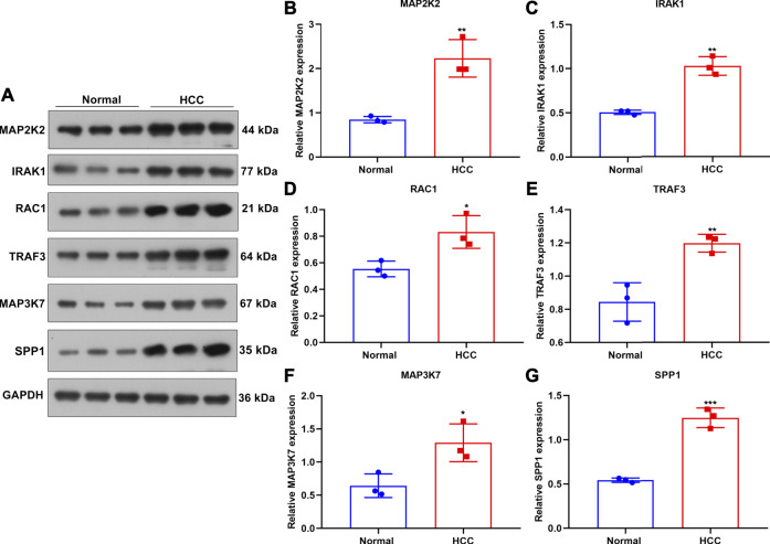
FIGURE 8.
Verification of the expression of genes in the TLR-based signature in HCC and normal tissue specimens. (A) Western blot for detecting MAP2K2, IRAK1, RAC1, TRAF, and MAP3K7 proteins in three paired HCC and normal tissues. (B–G) Quantification of the expression of (B) MAP2K2, (C) IRAK1, (D) RAC1, (E) TRAF, (F) MAP3K7, and (G) SPP1 proteins in three paired HCC and normal tissues. Comparisons between groups were evaluated with Student’s t tests. *p < 0.05; **p < 0.01; ***p < 0.001.

