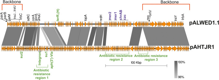
FIGURE 3.
Comparative linear map of plasmids pALWED1.1 and pAHTJR1. The location and polarity of genes and ORFs are shown with arrows. The extent of homologous regions is indicated in the dark gray shading. The backbones regions of plasmids are delimited by square red brackets and antibiotic resistance regions in pAHTJR1—by green. Antibiotic resistance genes are colored in green and genes of resistance to salts of heavy metals—in blue. Antibiotic resistance region 1 contains integron with the cassette genes arr-3- (rifamycine resistance) and aacA4 (aminoglycoside resistance) and the gene aph(3″)-V1a (aminoglycoside resistance). Antibiotic resistance region 2 contains the genes oxa58 (carbapenem resistance), the msrE and mphE (macrolide resistance) and the floR (phenicol resistance). Antibiotic resistance region 3 contains the aminoglycosides resistance genes aph(3″)-1b and aph(6) -1d and tetracycline resistance genes tet(Y) and tetR. Other genes: sdhC, succinate dehydrogenase, cytochrome b556 subunit; corA, magnesium and cobalt transport protein CorA; gorA, glutathione-disulfide reductase; umuC, DNA polymerase V subunit UmuC; topA, topoisomerase IA; nrdH, putative NrdH-redoxin family protein.

