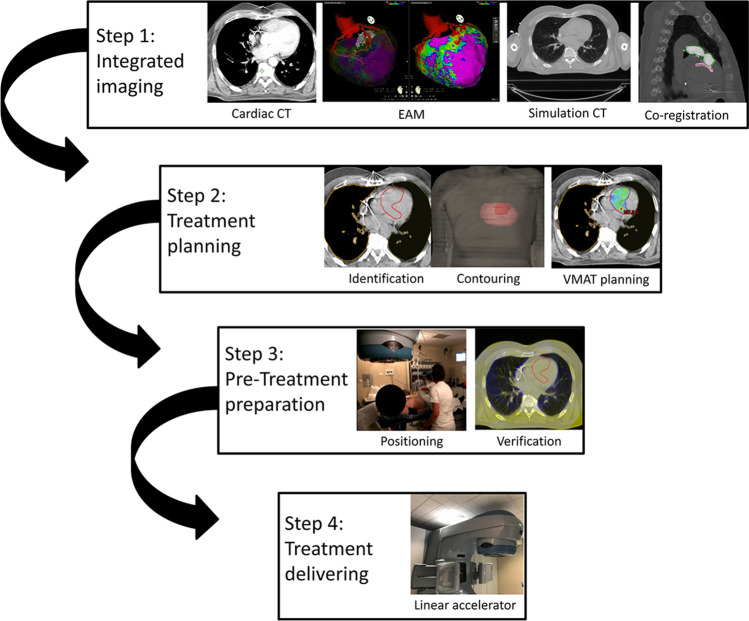
Fig. 1.
Interventional workflow. Step 1—cardiac CT data, integrated with endo-epicardial electroanatomical mapping, are merged with simulation “free-breathing” CT, acquired together with a “breathing-triggered” 4D-CT. Step 2—integrated “free-breathing” simulation-CT imaging is used as platform for the identification and contouring of the clinical target volume and of the organs at risk. The volumetric modulated arc therapy treatment plan is processed by the Eclipse RapidArc Planning System to deliver a single-fraction total dose of 25 Gy. Step 3—patient’s positioning setup is ensured by means of a vacuum immobilization cast on the treatment couch and is verified during the whole treatment process; two to three cone-beam CT scans are performed (image-guided radiotherapy), if necessary, for setup optimization. Step 4—the volumetric modulated arc therapy treatment is eventually delivered using the Varian Trilogy linear accelerator with the patient in the conscious state, lying down in a comfortable position in his/her immobilization cast. Abbreviation: CT, computed tomography

