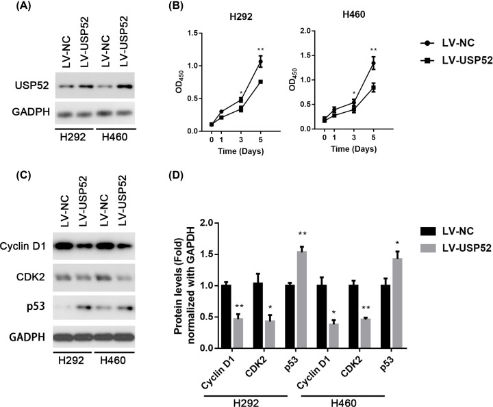
Figure 2. Overexpression of USP52 inhibits cell growth in NSCLC cells.
(A) H292 and H460 cells were stably infected with lentiviral LV-NC or LV-USP52, followed by Western blot against USP52. GAPDH was used as a loading control. (B) H292 and H460 cells stably infected with LV-NC or LV-USP52 were cultured for indicated time, followed by CCK-8 assay at days 0, 1, 3 or 5. (C) H292 and H460 cells were stably infected with l LV-NC or LV-USP52, followed by Western blot against CCND1, CDK2, p53 and GAPDH. (D) The relative abundance of CCND1, CDK2 and p53 in H292 and H460 from (C) were quantified by gray scanning after LV-USP52 plasmids transformed at 0, 1, 2, 4 μg. These experiments were repeated for three times. One-way ANOVA, *P<0.05, **P<0.01. NS, not significant.

