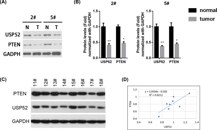
Figure 4. USP52 and PTEN were decreased in NSCLC tissues.
(A) Two representative pairs of fresh primary NSCLC tumor tissues (T) and individual normal paracancerous tissues (N) were prepared for Western blot against USP52 and PTEN. (B) The relative abundance of USP52 and PTEN in N and T from (A) were quantified by gray scanning as 2# and 5# patients, respectively. These experiments were repeated for three times. One-way ANOVA test, *P<0.05, **P<0.01. NS, not significant. (C) Eight representative pairs of fresh primary NSCLC tumor tissues were prepared for Western blot against USP52, PTEN and GAPDH. (D) Linear function was used to analyze the correlation between USP52 and PTEN expression, and fit linear function Y = 1.0034x-0.182, R2 = 0.6211.

