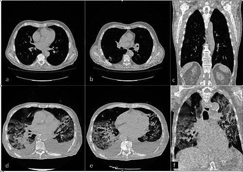
Figure 2.

a-f ) Chest CT of an 89-year-old man with ICU COVID-19 pneumonia (a). An axial and coronal CT image showed diffuse large regions of crazy-paving pattern with partial consolidation and bilateral pleural effusion. CT findings in a 54-year-old man with non-ICU COVID-19 pneumonia (b). An axial and coronal CT image showed bilateral multiple small regions of subpleural GGO and consolidation.
