
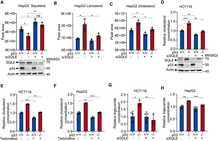
Figure 4. p53 inhibits cholesterol metabolism through SQLE.
-
A–CSqualene (A), lanosterol (B), and cholesterol (C) abundance in p53 +/+ and p53 −/− HepG2 cells transfected with control or SQLE siRNA for 48 h were determined via ultra‐high pressure liquid chromatography coupled to mass spectrometry (UHPLC‐MS). Protein levels were analyzed by Western blotting with specific antibodies (A bottom panel).
-
Dp53+/+ and p53−/− HCT116 cells were transfected with control or SQLE siRNA for 48 h as indicated. Relative cholesterol concentrations were examined as described in methods. Protein expression was determined by Western blot.
-
E, FCholesterol levels of HCT116 cells (E) and HepG2 cells (F) treated with or without 30 μm terbinafine for 72 h.
-
G, HTriglyceride levels in p53 +/+ and p53 −/− HCT116 cells (G) and HepG2 cells (H) treated with SQLE siRNA or control siRNA for 48 h.
Data information: (A–H), bars represent mean ± s.d., *P < 0.05; **P < 0.01; ***P < 0.001; n = 3 biologically independent samples; statistical significance was determined by two‐tailed unpaired t‐test.