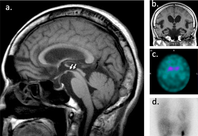
Figure 4.
Progressive supranuclear palsy with disproportionately enlarged subarachnoid space hydrocephalus (DESH). (A) Sagittal section of brain magnetic resonance imaging (MRI) in a progressive supranuclear palsy (PSP) patient showing midbrain atrophy with preservation of the pons (below white arrows) known as the “hummingbird” sign. (B) Coronal section of brain MRI showing “DESH” sign. Dopamine transporter imaging with [123I] N-ω-fluoropropyl-2β-carboxymethoxy-3β-(4-iodophenyl) nortropane (FP-CIT) single-photon emission computed tomography (SPECT) images (C) and 123I-meta iodobenzylguanidine (MIBG) cardiac scintigraphy (D). PSP patients have a significantly lower uptake of FP-CIT in the caudate nucleus and putamen than iNPH patients and normal individuals. In PD and LBD, there is nearly no MIBG uptake in the myocardium, while normal MIBG uptake in the myocardium is shown in PSP. iNPH – idiopathic normal pressure hydrocephalus; PD – Parkinson’s disease; LBD – Lewy body dementia.

