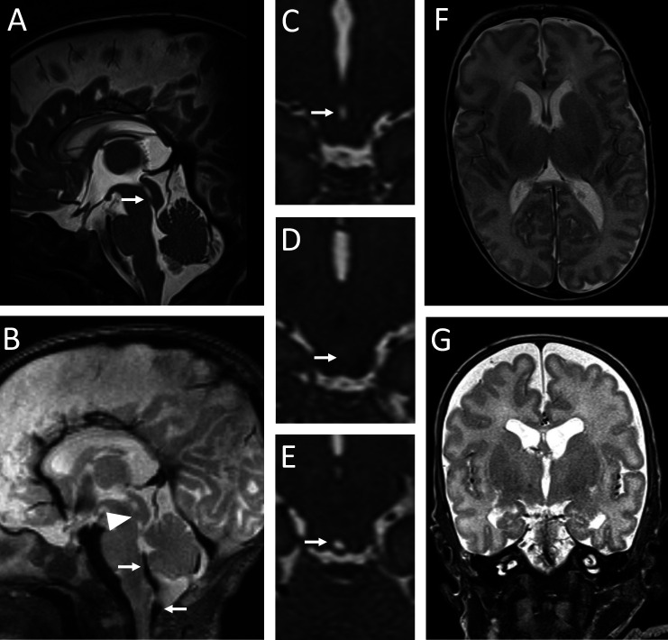
Figure 1.
A magnetic resonance (MR) exam in Patient 1 at term-equivalent age. A mediosagittal T2 ZOOMit section shows a membrane in the caudal half of the mesencephalic aqueduct (arrow in A). Artifacts of cerebrospinal fluid movement are visible in the area of the foramen of Magendie and cranio-cervical junction (arrows in B), but are absent from the mesencephalic aqueduct (arrowhead in B). An axial T2 ZOOMit section through the mesencephalic aqueduct cranial to the obstruction site shows a maintained aqueduct lumen (arrow in C). An axial T2 ZOOMit section through the aqueductal membrane shows a complete obstruction of the mesencephalic aqueduct (arrow in D). An axial T2 ZOOMit section through the mesencephalic aqueduct caudal to the obstruction site shows a maintained aqueduct lumen (arrow in E). Axial (F) and coronal (G) T2 sections through the brain parenchyma show normal volume of the lateral ventricles and third cerebral ventricle.

