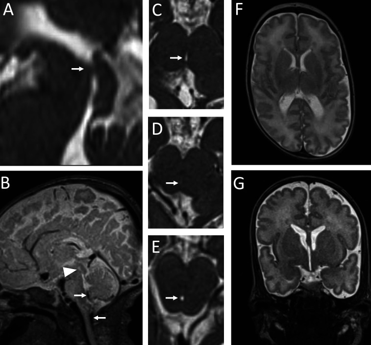
Figure 3.
A magnetic resonance (MR) exam in Patient 2 at term-equivalent age. A mediosagittal T2 ZOOMit section shows an obstruction in the cranial portion of the mesencephalic aqueduct (arrow in A). Artifacts of cerebrospinal fluid movement are visible in the area of the foramen of Magendie and the cranio-cervical junction (arrows in B), but are completely absent from the mesencephalic aqueduct (arrowhead in B). An axial T2 ZOOMit section through the mesencephalic aqueduct cranial to the obstruction site shows a maintained aqueduct lumen (arrow in C). An axial T2 ZOOMit section at the obstruction level shows a completely obstructed aqueduct lumen (arrow in D). An axial T2 ZOOMit section through the mesencephalic aqueduct caudal to the obstruction site shows a maintained aqueduct lumen (arrow in E). Axial (F) and coronal (G) T2 sections through the brain parenchyma show adequate volume of the lateral ventricles and third cerebral ventricle.

