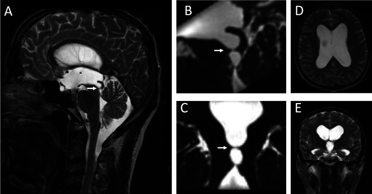
Figure 4.
A magnetic resonance (MR) exam in Patient 3 at the age of 56 years. A mediosagittal T2 sequence shows an aqueductal membrane obstructing the mesencephalic aqueduct (arrow in A). 3D T2 CISS sections showing the aqueductal membrane completely dividing the central part of the mesencephalic aqueduct in the sagittal (B) and coronal planes (C). Axial (D) and coronal (E) T2 sections through the brain parenchyma show an enlarged third ventricle and lateral ventricles without transependymal cerebrospinal fluid edema that would indicate increased intraventricular pressure.

