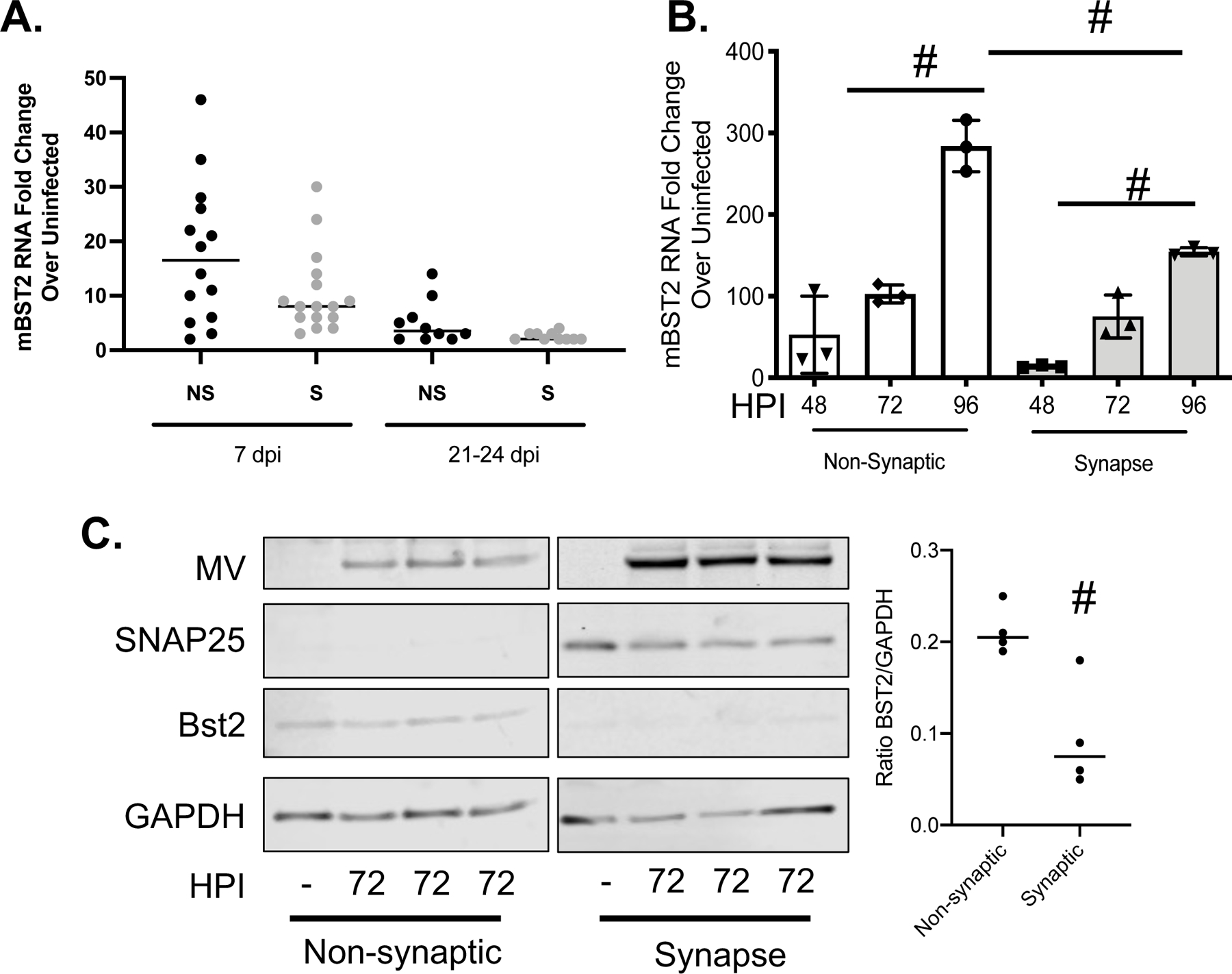
Figure 3: BST2 is not concentrated at the neuronal synapse during MV infection.

A) RNA collected from fractionated whole brain tissue was analyzed by RT-qPCR for murine BST2 (mBST2) RNA and normalized to cyclophilin. B/C) Primary NSE-CD46+ neurons were infected with MV at an MOI=1. Infected cells were collected at the indicated hours post infection followed by synaptosome purification. B) RNA was analyzed by RT-qPCR for murine BST2 (mBST2) RNA and normalized to cyclophilin B. Data represent the results of an experiment performed in triplicate and analyzed using the ΔΔCT method. C) Western blot analysis of protein collected at the indicated times post infection from either the pelleted synaptic or remaining fractions (non-synaptic). Blots were probed with antibodies against BST2, MV, SNAP25 (to indicate synaptic fraction purity) and an antibody to GAPDH (loading control). # p <0.05 Unpaired T test with equal standard deviations. Error bars represent SD.
