Figure 3.
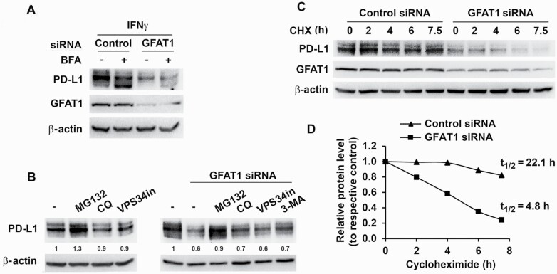
Knockdown of GFAT1 accelerates PD-L1 degradation by proteasomal pathway. (A) H2009 cells were transfected with control or GFAT1 siRNA for 24 h and then incubated with IFNγ for 6 h with or without prior brefeldin A (BFA, 5 µM) treatment for 30 min. (B) H2009 cells were treated with inhibitors [MG132, 1 µM; chloroquine (CQ), 20 µM; and 3-methyladenine (3-MA), 5 mM; left panel] or transfected with control or GFAT1 siRNA. Six hours post-transfection, media were refreshed and inhibitors (MG132, 1 µM; CQ, 20 µM; VPS34 inhibitor, 10 µM; and 3-MA, 5 mM) added (right panel). Total cell lysate was prepared 24 h later for western blot. Low concentration (1 µM) of MG132 was used to minimize cell death in this setting. (C and D) H2009 cells were transfected with control or GFAT1 siRNA for 24 h. Cycloheximide (CHX, 10 µM) was added to the cells at different time points. PD-L1 levels were detected by western blot. Band densities were quantified and normalized to loading control and the half-life (t1/2) of PD-L1 was calculated using Microsoft Excel equation with corresponding untreated control set as 1.

