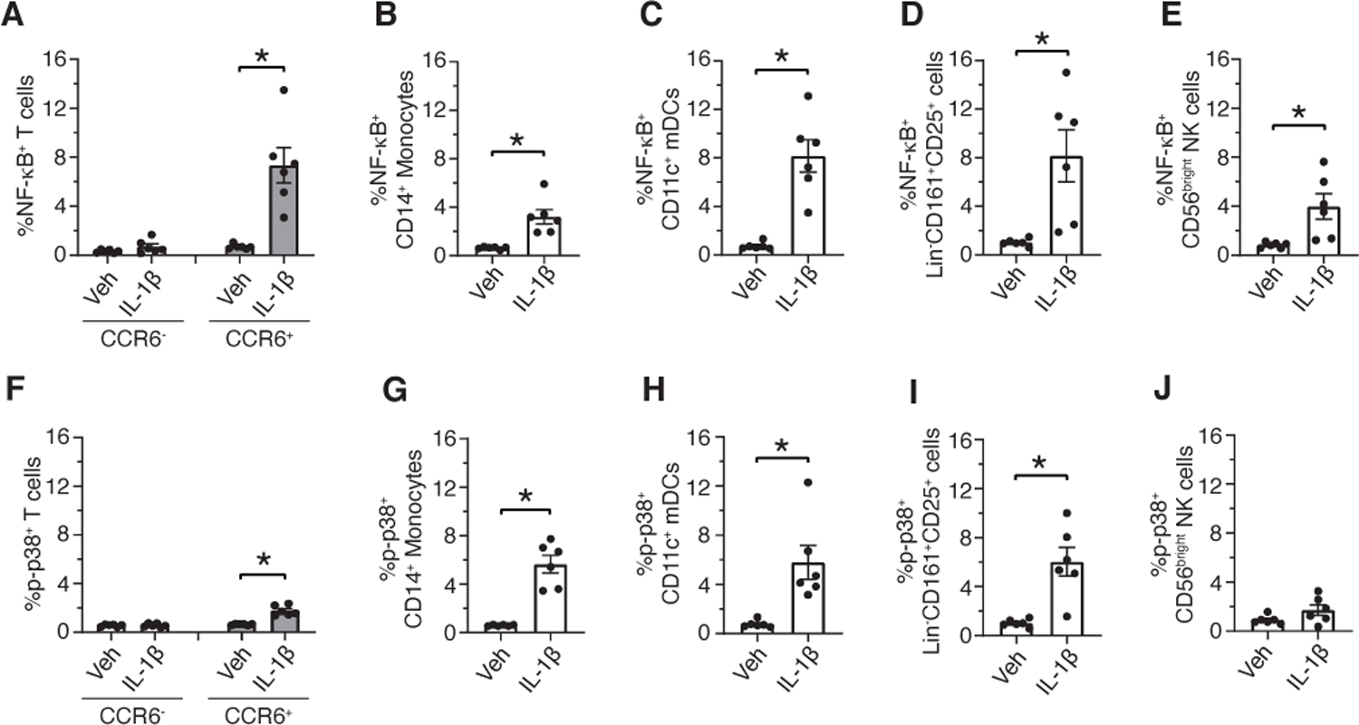
Fig 8. CyTOF analysis of an independent cohort identified IL-1β induced signaling activation in the same immune cell subtypes.

(A to J) PBMCs were incubated with vehicle (Veh) or IL-1β for 15 minutes. Cells were fixed, barcoded, and stained with cell surface and intracellular phosphoprotein antibodies. Debarcoded, normalized CyTOF data was analyzed in Flowjo. Frequency of p-NF-κB+ and p-p38+ CCR6+ T cells (A and F), p-NF-κB+ and p-p38+ CD14+ monocytes (B and G), p-NF-κB+ and p-p38+ CD11c+ mDCs (C and H), p-NF-κB+ and p-p38+ Lin-CD161+CD25+ cells (D and I) and p-NF-κB+ and p-p38+ CD56bright NK cells (E and J). Data shown are mean ± SEM. Wilcoxon signed-rank test; * p<0.05. N=6 donors for A-J.
