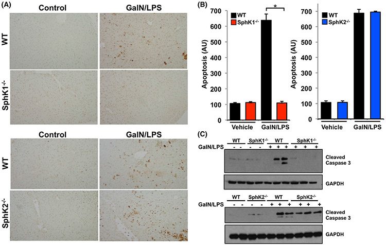
FIGURE 2.
SphK1 deletion protects from GalN/LPS-induced apoptosis. A-C, WT, SphK1−/−, and SphK2−/− mice were treated without or with GalN/LPS. (A,B) After 4 hours, liver sections were TUNEL stained to visualize apoptosis and quantified as described in Materials and Methods. Data are expressed as arbitrary units (AU) and are means ± SEM. n = 3. * P < .01 compared to WT. C, Proteins in liver lysates were separated by SDS-PAGE and analyzed by immunoblotting with antibody for cleaved caspase 3. Blots were re-probed with anti-GAPDH to show equal loading and transfer

