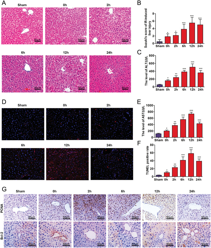
FIGURE 1.

Time‐dependent pathological changes resulting from hepatic IRI in mice. (A) Ischemia, necrosis, and edema as observed at different time points following IRI (scale bars = 50 μm). (B) Suzuki scores in liver IRI. (C and E) Serum ALT and AST levels of mice as a function of increasing reperfusion time. (D) Apoptosis as determined using TUNEL. (F) Analysis of TUNEL positive cells in the liver. (G) Expressions of PCNA and Bcl‐2 in liver as determined using immunocytochemistry (scale bars = 50 μm). * p < 0.05, ** p < 0.01, and *** p < 0.001 compared with sham group
