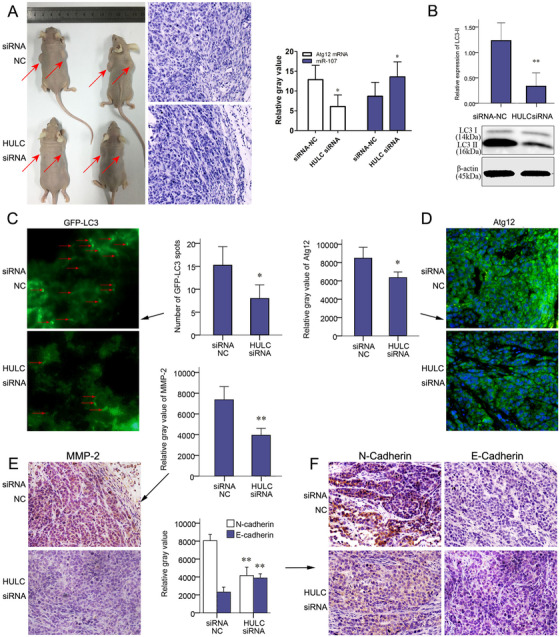
FIGURE 7.

HULC‐promoted invasion of subcutaneous planted tumors. After transfecting with a LV‐HULC siRNA or LV‐siRNA‐NC, SMMC‐7721 cells were implanted (n = 8) into 5‐week‐old nude mice subcutaneously. A, Tumors in situ (red arrows), H&E staining of para‐tumor tissues, and qRT‐PCR results of miR‐107 and Atg12 mRNA in xenograft tumors in siRNA NC and HULC siRNA groups. B, The expression of LC3 quantified by western blots analysis in siRNA NC and HULC siRNA groups. C, At the end of the 5th week, 25 µL mRFP‐GFP LC3 adenoviral virus (1×10¹⁰ PFU/mL) was injected into the xenograft tumors of both groups to detect autophagy. Forty‐eight hours later, tumors were harvested and studied. Green puncta of autophagosomes were captured by fluorescence microscope in frozen sections of tumors. D, Expressions of Atg12 were analyzed by IF staining on paraffin section. (E) Expressions of MMP2 and (F) N‐Cadherin and E‐cadherin were showed by IHC staining in HULC siRNA and siRNA‐NC groups (magnification, 200×; scale bars = 50 µm). * P < .05 and ** P < .01 (compared with the first group)
