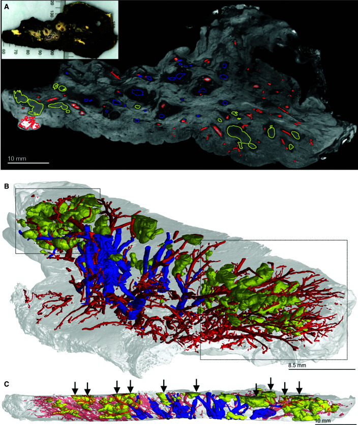
Figure 2.
Micro computed tomography and segmentation of a human tuberculosis lung slice. (A) Two-dimensional slice of micro computed tomography (52.0-μm resolution) of an upper lobe. Necrotic granulomas (yellow), bronchi and/or bronchioles (blue), and the vasculature (red) are outlined. The inset shows the gross image of the lung slice (see also Figure E1). (B) Lung slice in A at an angle to best illustrate the three-dimensional organization of granulomas (yellow), bronchi and/or bronchioles (blue), and the vasculature (red). Complex necrotic granulomas are oriented similarly to the airways and vasculature. The two boxed areas show the absence of airways in regions where granulomas predominate, suggesting replacement through bronchial obstruction. (C) Side view of A with transected granulomas indicated by vertical arrows. Scale bars: A and C, 10 mm; B, 8.5 mm. Reprinted by permission from Reference 41.

