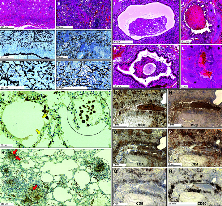
Figure 6.
Histopathology of the small airways of Mycobacterium tuberculosis–infected human lungs demonstrating bronchial obstruction. (A and B) Low-power magnification of hematoxylin and eosin staining of the lung parenchyma. (C and D) Low-power and (E and F) medium-power magnification of epithelial staining in the adluminal layer demonstrating alveolar consolidation (C and E, CK7; D and F, 3MPST). (G and H) Combined CD68 immunohistochemistry and Ziehl–Neelsen staining demonstrating the presence of macrophages and M. tuberculosis, respectively. Circled areas indicate alveoli filled with macrophages, and red arrows indicate giant cells. Yellow arrows indicate M. tuberculosis. (I–L) Hematoxylin and eosin staining of bronchial obstruction. Asterisks indicate cartilage. (M–R) Immunohistochemistry of myeloid cells and lymphocytes. For high-power images of boxed areas in M–R, see Figures E8 and E10 in the online supplement. Scale bars: A–D, F, and J, 500 μm; E, I, K, and H, 250 μm; G, 50 μm; L–Q, 1 mm; R, 2.5 mm. LCA = leukocyte common antigen; MPO = myeloperoxidase.

