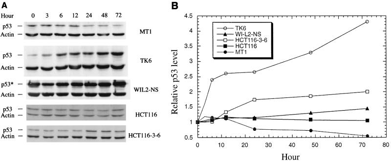
FIG. 7.
Expression of p53 in MMR-proficient and -deficient cells after treatment with AAAF. (A) Western blot analysis. After AAAF treatment, cells were cultured in fresh medium for the indicated times. Actin was used as an internal control, and equal amounts of protein were loaded for each sample. Both p53 protein and actin were detected by chemiluminescence on a Western blot by using antibodies against p53 and actin (Sigma), respectively. p53*, mutant p53 protein. (B) Relative amounts of p53. The p53 level at each point was normalized by its corresponding actin level as well as by the amount of p53 at time zero.

