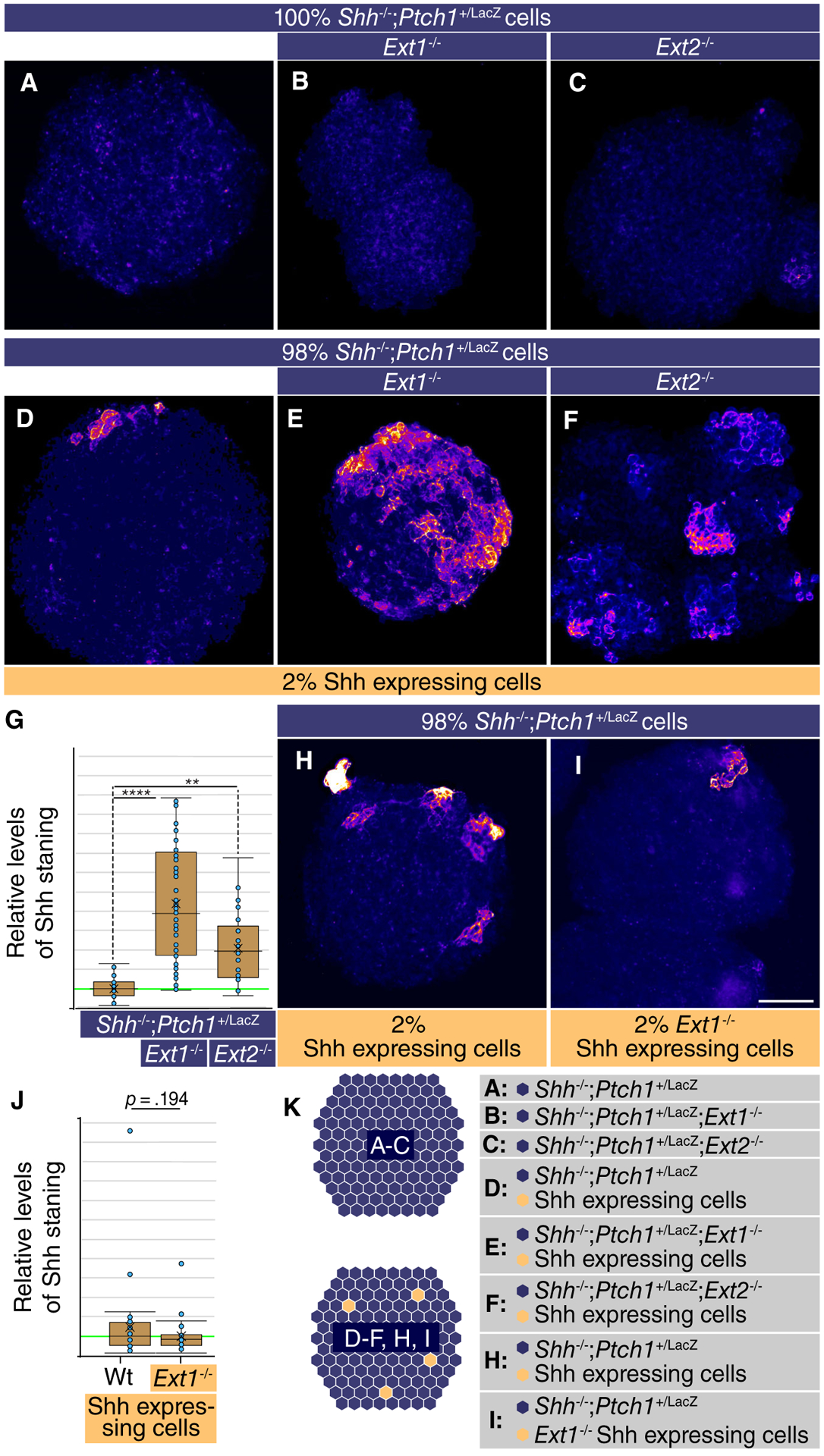
Figure 1.

Loss of Ext1/2 function in surrounding cells increases Sonic Hedgehog (Shh) accumulation around the source cells. (A–E, H, I): Neuralized embryoid bodies (nEBs) derived from mouse embryonic stem cell lines with the indicated composition and genotype. Extracellular Shh distribution was assayed by live staining after 4 days. nEBs derived from Shh−/−;Ptch1+/LacZ cells (A), Shh−/−;Ptch1+/LacZ;Ext1−/− cells (B), and Shh−/−;Ptch1+/LacZ;Ext2−/− cells (C), without embedded Shh-expressing cells. (D–F): Mosaic nEBs consisting of 98% either Shh−/−;Ptch1+/LacZ cells (D), Shh−/−;Ptch1+/LacZ;Ext1−/− cells (E), or Shh−/−;Ptch1+/LacZ;Ext2−/− cells (F), incorporating 2% Shh-expressing cells were live stained for Shh. (G): Quantification of (E)–(F). The level of Shh staining in positive areas was measured. One-way analysis of variance with post hoc Dunnett’s test; **, p < .01; ****, p < .0001. (H, I): Mosaic nEBs consisting either 2% wild-type Shh-expressing cells (H), or Ext1−/− Shh-expressing cells (I) and 98% Shh−/−;Ptch1+/LacZ cells were generated and assayed by live staining for extracellular Shh distribution. (J): Quantification of (H) and (I). (K): Diagram showing the experimental approach. Scale bar is 100 μm.
