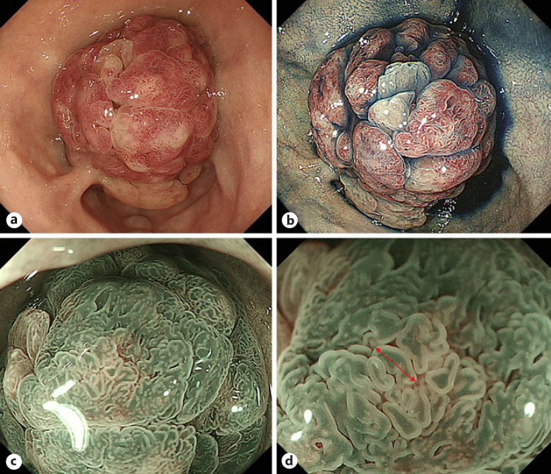
Fig. 1.
Endoscopic findings of a SNADET with the GP. a, b WLI shows a reddish pedunculated lesion in the first portion of the duodenum. The lobular/granular pattern was positive. c, d M-NBI reveals a dense pattern and DIP (red arrow). On the other hand, WOS and light blue crest are both negative. SNADET, superficial non-ampullary duodenal epithelial tumor; GP, gastric phenotype; M-NBI, magnifying endoscopy with narrow-band imaging; WOS, white opaque substance; WLI, white-light imaging; DIP, dilatation of the intervening part.

