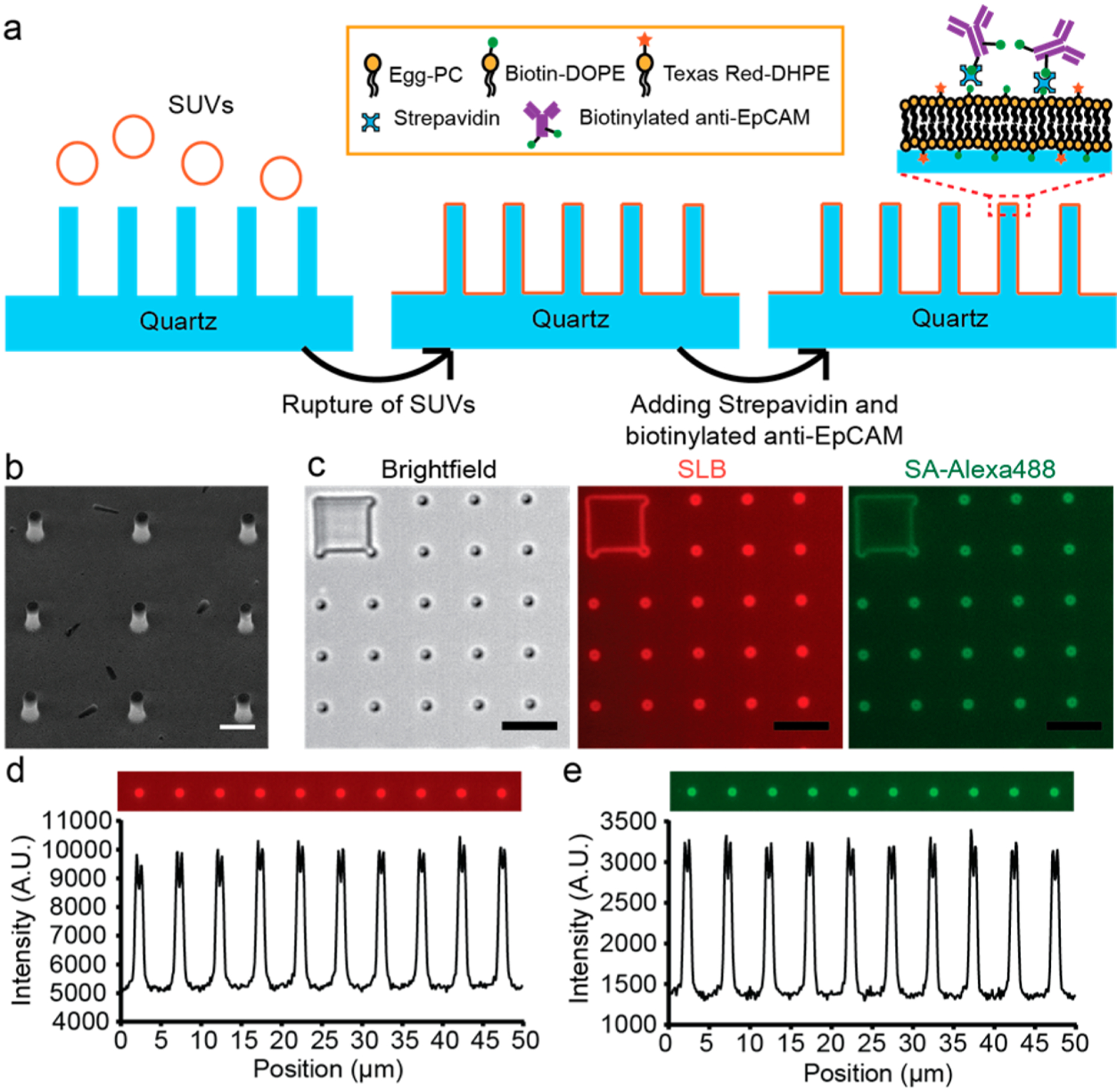
Figure 1.

Lipid-based functionalization of antibodies on quartz nanopillar arrays. (a) Schematic illustration of the lipid-based antibody functionalization procedure on a quartz nanopillar array. (b) Scanning electron microscopy (SEM) image of a quartz nanopillar array with 500 nm diameter, 3 μm spacing, and 1.3 μm height, tilting 30°. (Scale bar 1 μm.) (c) Bright field and fluorescence images of the supported lipid bilayer (SLB) and SA-Alexa488 on a quartz nanopillar array. The bright spots in the fluorescence images indicate that lipid bilayers also formed on the side wall of quartz nanopillars. (Scale bars 5 μm.) (d, e) Fluorescence intensity profile of SLB (d) and SA-Alexa488 (e) on quartz nanopillar arrays. The intensity shows that both SLB and SA-Alexa488 are distributed uniformly on each nanopillar.
