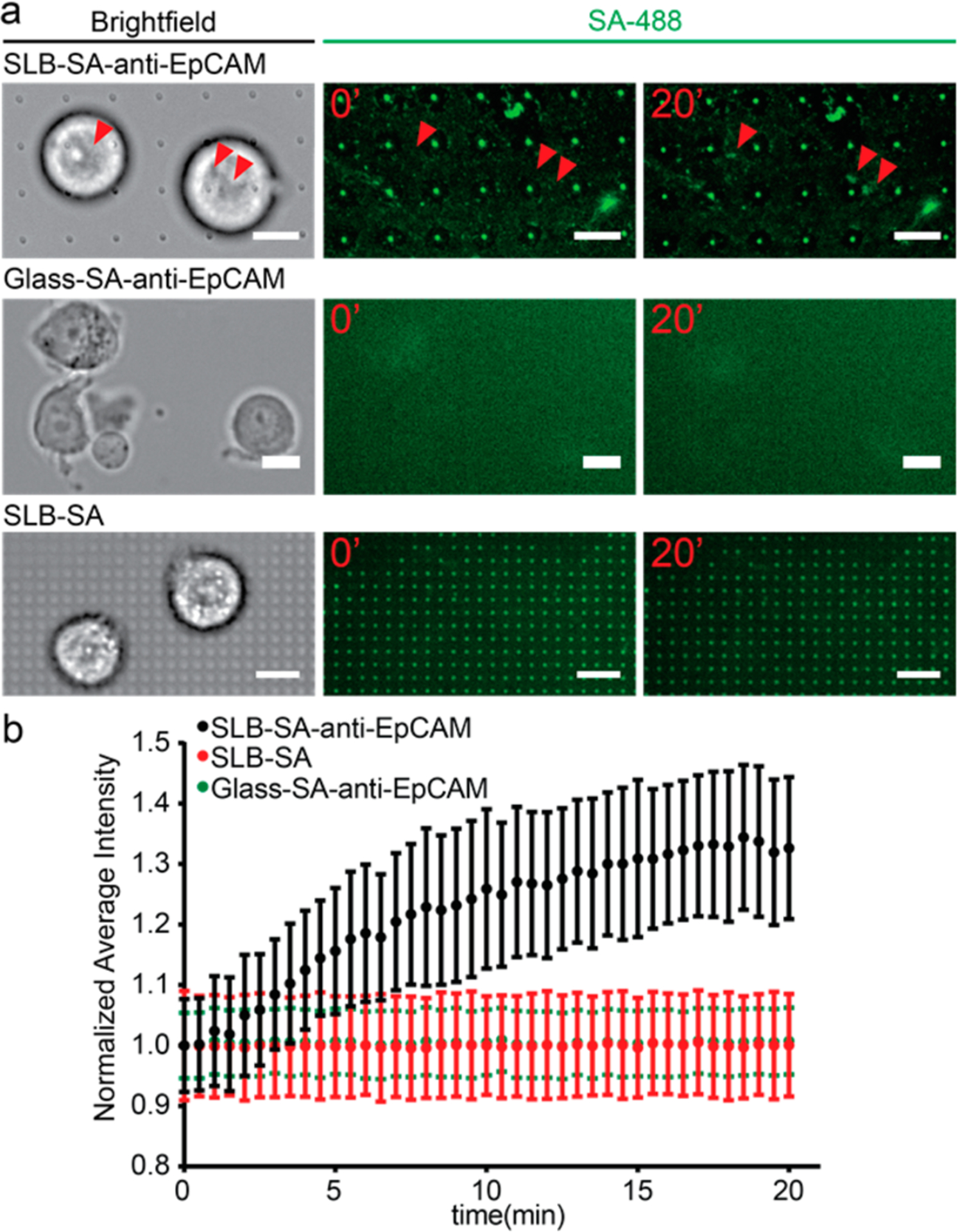
Figure 3.

Clustering of anti-EpCAM around captured MCF7 cells enabled by the antibody–antigen interaction and the fluidity of supported lipid bilayers. (a) MCF7 cells on different surfaces: supported lipid bilayer-coated quartz nanopillar arrays functionalized with SA-488 and anti-EpCAM (upper), bare glass coated with SA-488 and anti-EpCAM (middle), and supported lipid bilayer-coated quartz nanopillar arrays functionalized with SA-488 only (bottom). Only MCF7 on lipid bilayers with SA-488 and anti-EpCAM shows the clustering of SA-488 (red arrows). (Scale bars 20 μm.) (b) The average intensity change of SA-488 beneath the MCF7 cells on different surfaces. The average intensity of the cluster area increased about 30% in 20 min on SLB with SA-488/anti-EpCAM (black). However, no clustering of SA-488 was observed under MCF7 cells on surface-absorbed SA-488/anti-EpCAM (green) or the supported lipid bilayer with SA-488 only (red).
