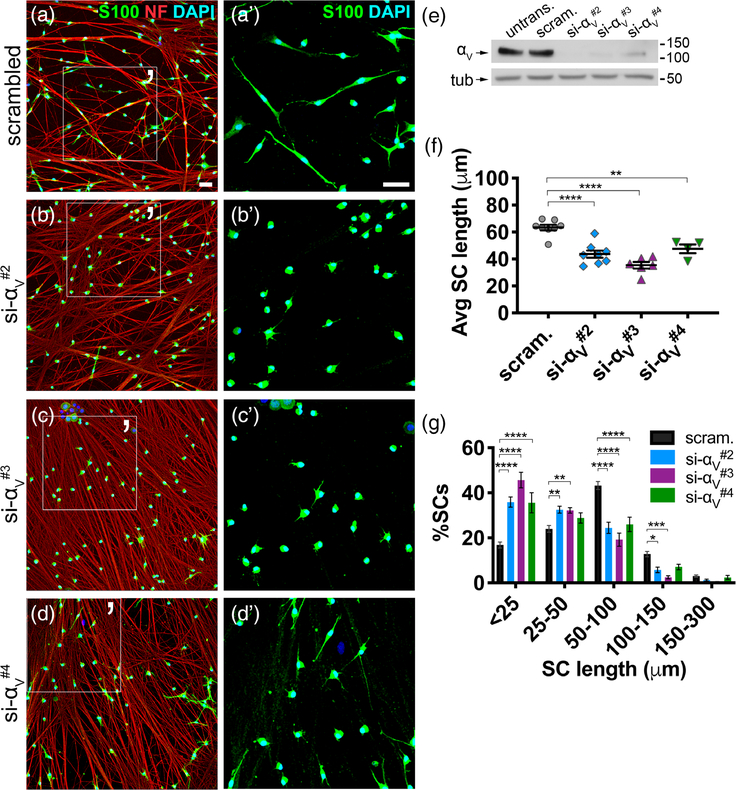
FIGURE 3.
Silencing the integrin subunit αV in SCs phenocopies the effect of RGD antagonists in SCs. (a–d) SCs were transfected with scrambled siRNA (scram., negative control) or one of three sequences targeting the integrin αV subunit (si-αV#2, -αV#3, -αV#4), then cocultured with untreated WT DRG neurons for 3.5 hr. Cultures were stained with S100 (green), neurofilament (NF) (red), and DAPI (blue) to label SCs, neurons, and nuclei, respectively. (a′–d′) Magnified area of (a–d). Scale bar is 50 μm in all panels. (e) Immunoblot of untransfected (untrans.), scrambled (scram), and si-αV-treated SCs to confirm αV protein knockdown. (f) Average (Avg) SC length in each cohort (student’s t test; scrambled: n = 8 replicates, 3,365 total cells quantified; si-αV#2: n = 8 replicates, 3,563 total cells quantified; si-αV#3: n = 6 replicates, 2,548 total cells quantified; si-αV#4: n = 4 replicates, 1,451 total cells quantified; SEM error bars). Compared to negative control, average length decreased in si-αV#2 (−31%), si-αV#3 (−44%), and si-αV#4 (−25%) cohorts. (g) SC lengths categorized into bins. Significantly more αV-silenced SCs were stunted (<25 μm), more had short processes (25–50 μm), and fewer lengthened (50–100 μm and 100–150 μm) compared to negative control (ordinary two-way analysis of variance (ANOVA), n = ≥4 coverslips, SEM error bars)

