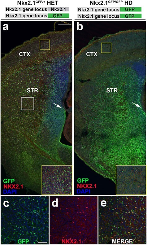
Figure 3 .

Residual GFP reporter retained in Nkx2.1 heterozygous and HD mice serves to fate map Nkx2.1-derived cells. (a,b) Low magnification E18.5 hemi-section of Nkx2.1GFP/+ heterozygous and Nkx2.1GFP/GFP HD mice. (a) Nkx2.1GFP/+ HET brain slice co-stained with NKX2.1 antibody and DAPI shows widely distributed precursor cells across neocortex and basal ganglia. Yellow boxed inset shows high magnification of GFP+ cells occupying neocortex with immature neuronal morphology. White boxed region shown in (c–e) reveal numerous GFP+ neuroblasts are immmunopositive for NKX2.1, consistent with normal NKX2.1 expression in heterozygous animals. In contrast, (b) Nkx2.1GFP/GFP HD mice reveal near complete absence of GFP+ cells from neocortex (yellow boxed inset) with concomitant increase in overall GFP expression in developing basal ganglia. Immunohistochemistry shows almost complete loss of NKX2.1 expression consistent with global knockout resulting from GFP insertion into both Nkx2.1 gene loci. HET—heterozygous and HD—homozygous deletion. Scale bars represent 200 μm (a,b) and 50 μm (c–e).
