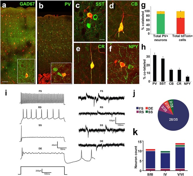
Figure 8 .

Neurochemical and functional analysis of Isl1-derived neurons shows fate bias towards fast-spiking cortical interneurons. (a) TdTomato+ neurons span all cortical layers and are largely co-positive with GAD67. (b inset, c–f) High magnification of cortical interneuron subtypes (b) parvalbumin, (c) somatostatin, (d) calbindin, (e) calretinin and (f) neuropeptide-Y co-labeled with tdTomato+ in cerebral cortex. (g) Left bar graph of co-positivity of total parvalbumin+ cells shows 12.1% ± 0.7 co-labeled (PV+/tdTomato+, yellow) cells and 87.9% ± 0.7 (PV only, green) cells. Right bar graph of co-positivity of total tdTomato+ cells shows 29.1% ± 2.7 (PV+/tdTomato+, yellow) cells and 70.9% ± 2.7 (tdTomato+ only, red) cells. (h) Bar graph shows % co-label of cortical tdTomato+ cells with all analyzed neurochemical markers shown in (b–f). (i) Electrophysiological traces of examples interneuron subtypes. Population data of recorded cells indicates the majority are fast spiking. Isl1-derived neurons exhibit 4 different electrophysiological properties. Current-clamp (left) and synaptic (right) traces for fast spiking (FS), regular spiking (RS), delayed excitation (DE), and single spiking (SS) interneurons. (j) Pie chart displaying the contribution of each electrophysiological property noted for Isl1-derived neurons. (k) Histogram summarizing the cortical distribution of each electrophysiological subtype of recorded Isl1-derived neurons. Scale bars represent 100 μm (a,b) and 20 μm (c–f).
