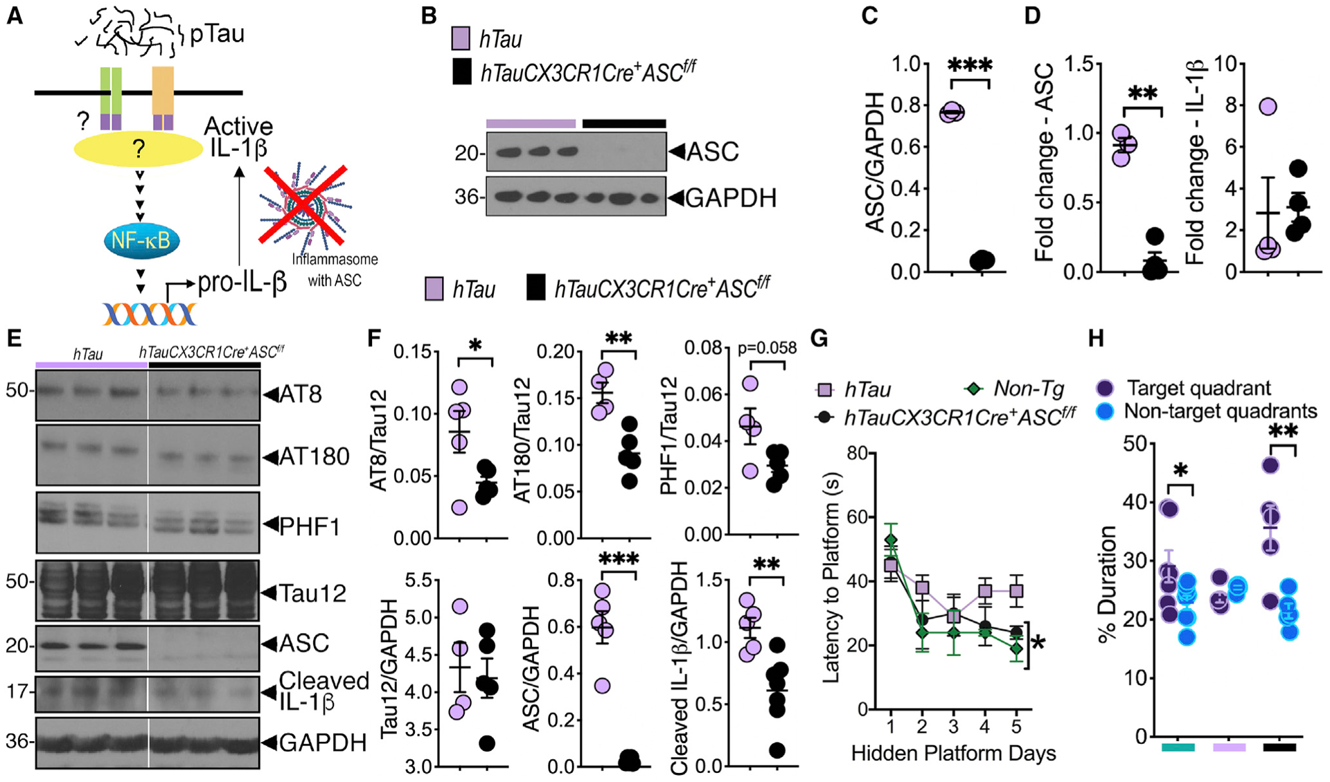
Figure 5. Myeloid cell-restricted deletion of ASC reduces IL-1β maturation and tau pathology and improves cognitive function in hTau mouse model of tauopathy.

(A) Working model shows potential mechanism of pTau-induced ASC-inflammasome activation/IL-1β maturation, which can be blocked by myeloid/microglial cell-specific deletion of ASC.
(B and C) Western blot analysis and quantification shows complete deletion of ASC in purified microglia from hTauCX3CR1Cre+ASCf/f mice.
(D) Microglia-restricted deletion of ASC shows significant reduction of ASC, but not IL-1β, mRNA expression in the brains of hTauCX3CR1Cre+ASCf/f mice compared to hTau controls.
(E and F) Western blot analysis and quantification of 8-month-old hTauCX3CR1Cre+ASCf/f mice shows reduced tau pathology (on AT8, AT180, and PHF1 sites), cleaved (activated) IL-1β, and ASC in the hippocampus compared to age-matched hTau controls.
(G) Morris water maze (MWM) analysis shows improved spatial learning to reach hidden platform by day 5 in 8-month-old hTauCX3CR1Cre+ASCf/f mice that was comparable to non-transgenic mice. This learning measure was impaired in age-matched hTau mice.
(H) Probe trial analysis of MWM test shows hTauCX3CR1Cre+ASCf/f mice spent significantly more time in the target quadrant compared to average duration in all other non-target quadrants.
Data displayed as mean ± SEM, *p < 0.05, **p < 0.01, ***p < 0.005, unpaired Student’s t test, n = 3–5 (B–D), n = 4–7 (E and F), or two-way ANOVA followed by Tukey’s multiple comparison test, n = 5–10 (G), multiple t test, n = 5–10 (H).
See also Figures S7F and S7G and Data S1.
